CYMOPLIVE – Cyto-Motility and Cyto-Plasticity in Vitro Live-Cell Assay

About the technology

Cymoplive is a platform that allows us to study cells in action and how they smartly tackle obstacles in our body. Cell movement or motility governs how cells migrate, invade, repair injuries, and develop tissues and organs. These processes are critical for a healthy, functioning body. Cell plasticity, in the form of individual or collective cell migration, has long-standing consequences and can help identify diseases and how much they have progressed. In the Cymoplive platform, cells are placed in a 3D maze where they need to overcome designed obstacles as they move through the maze. Cymoplive has a live-imaging friendly format compatible with all optical microscopes. The complexity of the maze can be customized based on the experimental setup and what is to be studied.

Value proposition

  • Live-imaging of process
  • Intelligence of cell movement can be studied
  • Mimics tissue environment with the 3D patterned obstacles matrix
  • Customisable physical environment
  • Customisable chemical environment
Image: Cymoplive is a unique cell migration assay platform that contains 3D obstacles in a solid hydrogel environment to study cell motility in a physiologically and functionally relevant environment.

Resources and partners

  • UiT Nanoscopy group
  • EU H2020-funded FET-Open RIA project OrganVision (EIC Pathfinder)
  • Proof of concept in a small-scale lab setting has been demonstrated
  • Patent application filed

Opportunities for Collaboration

We are currently looking for industry partners or potential users who finds interest in our technology. We are open for working together on the development of Cymoplive to accommodate your needs.

Image: The image shows how embryonic fibroblast cells are moving in the maze by changing their direction as they encounter obstacles on their path from high cell density to a low cell density region. In phase microscope, the morphological features of the cells can be observed and evaluated labelfree. In fluorescence microscopy, the molecular changes due to cells’ motility in the maze can be measured. Here we see how the actin cytoskeleton is modified to maneuver cells along the maze path.

Collaboration partners

Universitetet i Tromsø

Contact information

Foto: Marius Fiskum

Ingrid Skjæveland

Forretningsutvikler | Tech Transfer

ingrid@norinnova.no
Tlf. 991 57 143

Les om andre prosjekter:

mSpider

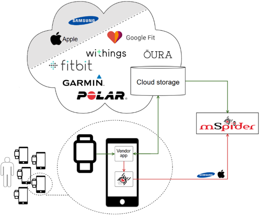
Motivating continous Sharing of Physical activity using non-Intrusive Data Extraction methods Retro- and prospectively About the technology Data collection of physical data …

Thyroid Assist

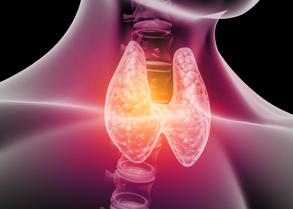
– A novel decision support aid tool (DST) for optimal levothyroxine dosage after thyroidectomy About the technology Levothyroxine is a necessary synthetic …

About the technology New and advanced label-free and super resolution microscopes and nanoscopes have emerged, and their characterization is becoming more demanding. …

Nanospacer

A powerful tool for nanoscopic size analysis and detection About the technology Nanospacer is a specially designed microscope coverslip which makes it …

A tool that predicts ice amount and distribution on marine structures About the technology Due to climate changes the Arctic marine territories …

CYMOPLIVE – Cyto-Motility and Cyto-Plasticity in Vitro Live-Cell Assay About the technology Cymoplive is a platform that allows us to study cells …